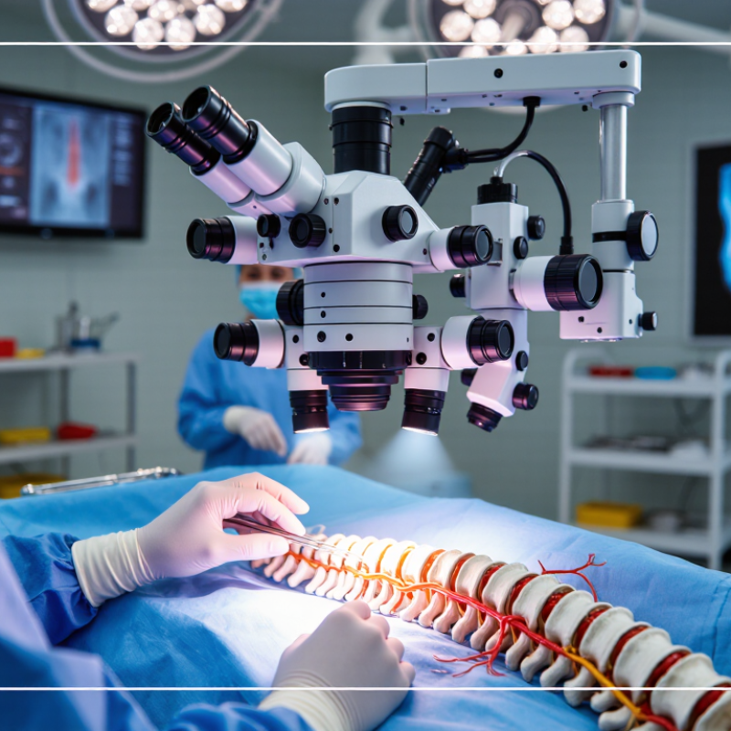
A surgical microscope is most often used when a surgeon needs a magnified, brightly lit view of small nerves and blood vessels. In lumbar microdiscectomy, it helps guide removal of disc material pressing on a nerve root. It can also assist with lumbar decompression, including laminotomy, and selected cervical procedures such as foraminotomy or anterior discectomy. Not every case needs one. The choice depends on the anatomy, procedure, and surgeon’s training.
Microscopes may also be used in delicate operations around the spinal cord, such as removing certain intradural tumors. During spinal fusion, they can aid the decompression portion, but they are not required for every fusion.
AHRQ’s Healthcare Cost and Utilization Project analysis found that U.S. inpatient spinal fusion procedures increased about 70% from 2001 to 2011. That figure describes fusion volume, not microscope use; national procedure data do not establish how often microscopes are used.
Still, it shows how frequently surgeons may encounter fusion-related work.
A microscope offers detail, not certainty.
Surgeons must still protect neural tissue, manage bleeding, and judge what can safely be removed.